D-MICRO HIGHLY IMPROVES MULTI-PHOTON MICROSCOPY
WITH EASY-TO-USE FEMTOSECOND LASER TECHNOLOGY
Image quality in multi-photon microscopy depends drastically on the compression of the laser pulses at the sample. Every optical component in a microscope introduces temporal dispersion that increases the laser pulse duration.
Ensuring optimum pulse compression at the sample is not an easy task, and is harder to do for shorter pulses.
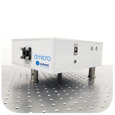
The d-micro comprises two COMPACT modules:
– the COMPRESSOR, which allows pre-compensating the dispersion introduced by the microscope
– the MEASURING HEAD, which measures the pulse directly at the sample position.
Sphere Ultrafast Photonics can install the d-micro system on a microscope already equipped with a femtosecond laser, or can provide a complete ultra-broadband femtosecond laser and bundled d-micro system, with guaranteed pulse duration below 10 fs at the sample plane.
Measurement examples
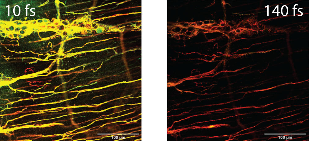
MULTI-FLUOROPHORE EXCITATION

At the same average power level in the sample plane, the fluorescence intensity produced by compressed broadband pulses was 2 – 3 times brighter than for long-pulse Ti:Sa pulses. When normalised against GFP intensity, compressed broad- spectrum pulses generated 10% more signal in the blue and red channels compared to uncompressed Ti:Sa laser pulses at 810 nm. These data support the superior imaging capabilities of compressed few-cycle laser pulses in 2p microscopy of biological samples.
For more details please check:
A. Manickavasagam, P. Fendel, M. Miranda,
P. T. Guerreiro, H Crespo, M. Renshaw, K. I. Anderson, “Multiphoton Excitation Of Biological Samples With Fourier- Limited, Broad-Spectrum, Ultrashort Laser Pulses “, Focus On Microscopy Congress 2019.
Credits: Kurt Anderson, Francis Crick Institute / Thorlabs / Sphere
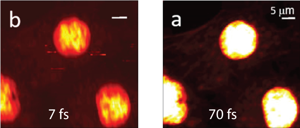
SyncRGB:FLIM

SyncRGB imaging using a 7 fs source. Multi-photon intensity images of the same sample areas taken with the compressed 70 fs Ti:Sapphire laser (a) and with the broadband 7 fs laser source (b). For both lasers, a filter configurations was chosen, a 685 nm short pass filter that transmits the emission wavelength ranges of all three chromophores. Scale bar in (a) is 5 μm and is used for all images and a pixel dwell time of 20 ms for all images. (a-b) are 100 × 100 pixels = 200 s/scan while (c-d) are 32 × 32 pixels = 20.5 s/scan.
For more details please check:
C. Maibohm, F. Silva, E. Figueiras, P. T. Guerreiro, M. Brito, R. Romero, H. Crespo, and J. B. Nieder, “SyncRGB-FLIM: synchronous fluorescence imaging of red, green and blue dyes enabled by ultra-broadband few-cycle laser excitation and fluorescence lifetime detection,” Biomed. Opt. Express 10, 1891-1904 (2019).
https://www.osapublishing.org/boe/abstract.cfm?uri=boe-10-4-1891
Technical Specifications
| d-micro US | d-micro S | d-micro LP R | d-micro LP NIR | d-micro LP 1.5 | |
| Wavelength range | 600-1100nm | 700-1400nm | 600-1100nm | 700-1400nm | 1500-1700nm |
| Pulse duration (FTL) | 5fs to 20fs | 2.5fs to 60 fs | 80fs to 140fs | 60fs to 200fs | |
| Dispersion Compensation Range | 0 to -4000fs2 | 0 to -8000fs2 | 0 to -300000fs2 | 0 to -20000fs2 | |
| Repetition rate | 1 kHz and above (a) | ||||
| Input polarization | Linear | ||||
| Max input aperture | 20 mm | ||||
| Required input energy | >100 pJ @ 80 MHz >1 µJ @ 1 kHz |
||||
| Compression module dimensions (WxLxH) | 317 x 336 x 97 mm | 250 x 250 x 100 mm | |||
| Measuring head dimensions (WxLxH) | 182 x 336 x 97 mm | 57 x 57 x 116mm | |||
(a) Lower repetition rates possible with external synch option
Talk to us for different wavelength range, chirp range, input aperture, and other


